October 21, 2024
Digital X-Rays
In modern dentistry, digital X-rays have revolutionized the way dental professionals diagnose and treat oral health issues. One of the most significant advantages of digital X-ray technology is its ability to reduce radiation exposure for patients. This blog will explore how digital X-rays work, their benefits over traditional film X-rays, and why they are a safer option for dental imaging.
Digital X-Rays: A Brief Overview
Digital X-rays are a form of radiography that utilizes digital sensors instead of traditional film to capture images of the teeth, gums, and surrounding structures. The images are then processed and displayed on a computer screen almost instantaneously. This technology not only streamlines the imaging process but also enhances the overall patient experience.
How Do Digital X-Rays Work?
- Sensor Technology: Digital X-rays use electronic sensors to capture the X-ray image, which is then converted into a digital format. These sensors are more sensitive to radiation compared to traditional film.
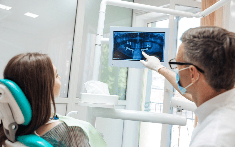
- Instant Image Display: Once the X-ray is taken, the image appears on the screen within seconds, allowing the dentist to analyze it immediately.
- Enhanced Image Quality: The digital format allows for image enhancement and manipulation, enabling better diagnosis.
Radiation Exposure: Understand The Risks
The Importance of Reducing Radiation
Radiation exposure is a significant concern in dental imaging. While the levels of radiation from X-rays are generally low, minimizing exposure is crucial, especially for certain populations, including:
- Children: Their developing tissues are more sensitive to radiation.
- Pregnant Women: Fetal exposure to radiation should be avoided when possible.
- Patients with Multiple X-rays: Individuals requiring frequent dental imaging are at a higher cumulative risk.
Traditional X-Rays or Digital X-Rays? Comparison Between These Two
Traditional film X-rays involve more radiation exposure compared to their digital counterparts. Here’s a comparison of the two:
- Radiation Exposure: Traditional X-rays have a higher radiation exposure, while digital X-rays have a lower radiation exposure.
- Image Processing Time: Traditional X-rays take longer to process, whereas digital X-rays provide instantaneous image results.
- Image Quality: The quality of traditional X-ray images is limited, while digital X-rays offer high-resolution images.
- Storage & Retrieval: Traditional X-rays require physical storage, while digital X-rays can be stored digitally.
- Environmental Impact: Traditional X-rays generate chemical waste, while digital X-rays produce minimal waste.
How Do Digital X-Rays Reduce Radiation Exposure?
1. High Sensitivity
Digital sensors are more sensitive to radiation than traditional film, allowing for the same diagnostic quality with significantly lower doses. This means that dentists can obtain high-quality images with reduced radiation levels.
2. Optimized Imaging Techniques
- Image Enhancement: Digital X-rays allow for immediate enhancement of images. Dentists can adjust contrast and brightness on the screen, enabling them to see details without needing multiple exposures.
- Selective Imaging: Dentists can focus on specific areas of concern, reducing the need for full-mouth X-rays, which would expose the patient to more radiation.
3. Lower Need for Retakes
With traditional film X-rays, retakes are often necessary due to poor image quality, positioning errors, or other factors. Digital X-rays significantly reduce the likelihood of retakes, further lowering overall radiation exposure. Some key points include:
- Immediate Feedback: The dentist can view the X-ray immediately and determine if a retake is necessary, minimizing the time spent in exposure.
- Precise Positioning: Digital X-ray systems often have features that assist with positioning, reducing the chances of image errors.
4. Advanced Imaging Options
Digital X-rays can integrate with advanced imaging technologies, such as Cone Beam Computed Tomography (CBCT), which provides three-dimensional images with reduced radiation doses compared to traditional 3D imaging methods.
Benefits of Digital X-Rays Beyond Radiation Reduction
While reducing radiation exposure is a significant advantage of digital X-rays, they offer several additional benefits, including:
Enhanced Patients’ Experience
- Quick Processing: Patients spend less time in the dental chair as images are available almost instantly.
- Comfort: Digital sensors are generally smaller and more comfortable than traditional film, leading to a better overall experience for patients.
Better Diagnosis and Treatment Planning
- Higher Resolution: Digital images provide greater detail, helping dentists diagnose issues more accurately.
- Ease of Sharing: Digital images can be easily shared with other specialists or stored in electronic health records, facilitating comprehensive care.
Environmentally Friendly
- Less Waste: Digital X-rays eliminate the need for film, chemicals, and physical storage, contributing to a more sustainable dental practice.
Digital X-rays represent a significant advancement in dental imaging, offering a host of benefits, particularly in reducing radiation exposure for patients. By utilizing more sensitive sensors and optimizing imaging techniques, dental professionals can provide safer, quicker, and more effective care.
As technology continues to evolve, the use of digital X-rays will likely become even more integrated into routine dental practices, ensuring that patient safety remains a top priority.
For anyone concerned about radiation exposure during dental visits, discussing digital X-ray options with your dentist can lead to a safer and more effective dental experience.
Embrace the future of dental imaging and enjoy the peace of mind that comes with knowing you are receiving care that prioritizes your health and safety.
Recent Posts

Feb. 02, 2026
Can Dr. Modjeski Help Me Get a Date-Ready Smile in Just One Visit?

Dec. 03, 2025
Fixing Chipped or Gapped Teeth: Can Porcelain Veneers Solve Multiple Smile Problems at Once?
Nov. 03, 2025
Fixing a Crossbite: Functional and Aesthetic Correction with Invisalign in Yorkville, IL

Oct. 03, 2025
The Clock is Ticking: 5 Essential Steps to Take Before You “Lose-It” on Your Dental Insurance Benefits
Sep. 05, 2025
The Longevity of Dental Implants: A Long-Term Solution for Yorkville Residents
Catagories
- cosmetic dentist in Yorkville (0)
- Cosmetic Dentistry (5)
- Crooked Teeth (1)
- Crowns and bridges (1)
- Dental Implants (18)
- Dentist in Yorkville (4)
- Dentures (4)
- Digital X-Rays (1)
- Emergency Dentistry (4)
- Invisalign (7)
- Oral Care (8)
- Oral Surgery (1)
- Periodontal Therapy (3)
- Porcelain Veneers (6)
- Prairie Garden Dental (44)
- Root Canal Treatment (4)
- Sleep apnea (4)
- Sumner Dental Group (0)
- Teeth Whitening (7)
- tooth extraction (3)
- Uncategorized (6)
- Veneers (2)